Optogenetics wins the Nobel – one neuroscience breakthrough in a line of technological innovations h
Neuroscientific advances follow a pattern of visualizing the brain, measuring its function and then controlling it. Optogenetics may lead to a new era of medical treatment.

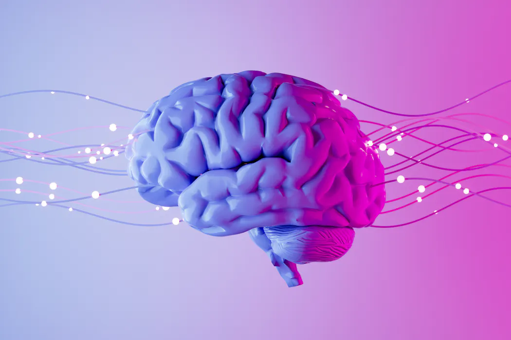
The brain is unbelievably complicated, which is both daunting and fascinating for neuroscientists like me. In the space of a cubic millimeter – about the size of a single poppy seed – there are tens of thousands of neurons and other brain support cells, all influencing each other’s function.
How do neuroscientists make sense of this complexity? And how did they get to the point where they could simply shine different colors of light on the brain to control neuron activity?
The answer is one step at a time, with a lot of help from technology.
Revisiting some of the technological advances that have supported researchers’ current understanding of how the brain works can point to where the field of neuroscience might be going next. There is a rough pattern to the technological advances that have led neuroscience to the present day: visualize the brain, measure its function and then control it.

Using light to influence neuronal function, known as optogenetics, allows neuroscientists to manipulate what neurons do millisecond by millisecond. The researchers whose work led to this technique won the 2026 Nobel Prize in physiology or medicine.
The level of control afforded by optogenetics suggests that the ability to therapeutically adjust brain activity in ways that previously sounded like fantasy could be on the horizon, including restoring eyesight, reducing pain and treating diseases like epilepsy and mental health disorders.
Visualizing the brain
Dollops of color and magnification are how neuroscientists came to know what neurons look like and how they connect to each other.
In the late 1800s, Spanish neuroscientist and artist Santiago Ramon y Cajal painted what he saw through the microscope. He looked at samples of brain tissue stained with silver to make individual neurons visible, a technique developed by the Italian neuroscientist Camillo Golgi.
As he painted, Cajal wondered about how neurons might influence each other through their connections. The ideas he developed form the foundation of how the brain learns and remembers. The work of these scientists won them the 1906 Nobel Prize in physiology or medicine.
In the 1970s, researchers developed another version of applying color to the brain called anterograde and retrograde tracing. These methods let neuroscientists map different pathways between neurons in the brain.
Today, the brain can be visualized in much finer detail using technologies such as electron microscopy, which involves firing a stream of electrons at an object to develop an image at atomic resolution, and expansion microscopy, which uses the same type of gel inside baby diapers to physically enlarge a tissue sample to visualize each cell and its connections. These techniques, combined with artificial intelligence algorithms, are what allow scientists to see the complexity within a cubic millimeter of brain tissue.
Magnetic resonance imaging has also profoundly influenced how researchers understand the brain. Functional MRI lets neuroscientists noninvasively eavesdrop on brain activity by measuring blood flow across the entire brain. In 2003, the Nobel Prize in physiology or medicine was awarded to the development of MRI techniques that make functional MRI possible: fast imaging sequences and spatial encoding.
All of these visualization and magnification methods form the field of connectomics: mapping all the connections between neurons.
Measuring neuronal activity
On top of visualizing the brain’s structures and layout, the ability to manipulate electricity has allowed researchers to learn how neurons communicate.
Neuroscientists Alan Hodgkin and Andrew Huxley won the 1963 Nobel Prize in physiology or medicine for their work on capturing and measuring the electrical impulses that neurons create. Neurons propagate these action potentials to communicate with each other.
Today, the probes that neuroscientists use to measure the electrical activity of neurons can listen in on the activity of hundreds of neurons from different brain areas at the same time.
Research using simultaneous recording methods is upending scientists’ understanding of how the brain works: A series of studies published in 2025 showed that while an animal is making a decision, 95% of the brain is actively involved in that process, not just neurons in brain areas typically associated with decision-making.
Controlling neurons
The ability to control the activity of specific neurons has been around since the late 1980s when French neurosurgeon Alim-Louis Benabid developed the technique of deep brain stimulation to help ameliorate the symptoms of Parkinson’s disease.
Deep brain stimulation acts like a pacemaker for brain activity. Today, researchers are studying the potential use of this technique to relieve symptoms of mental health disorders, including obsessive compulsive disorder, major depression and binge eating, to name a few.
While the results can be dramatic, deep brain stimulation is a relatively crude method for controlling neuronal activity because the implanted electrode influences all cells nearby. But researchers are developing more precise and higher resolution techniques to both study and adjust brain function.
One of the coolest things about optogenetic techniques is that they may also help treat disease. Researchers are currently testing whether optogenetics can alleviate symptoms or enhance the treatment of vision loss, Alzheimer’s disease, Parkinson disease epilepsy, multiple sclerosis, diabetes, cancer and more.
Neuroscience goes viral
Neurons don’t naturally respond to the lights used in optogenetics. So neuroscientists must instruct them to renovate their cell walls so that some of their proteins are responsive to light. Scientists do this by sending instructions directly to neurons via a harmless virus.
Optogenetics is not the only neuroscience technique that leverages viruses to deliver instructions to neurons. So does a method called designer receptors exclusively activated by designer drugs, or DREADDs for short.
Like optogenetics, DREADDs give neuroscientists the ability to control neuronal activity. While optogenetics uses light to zero in on a specific group of neurons to control their activity, DREADDs does this chemically – like a lock and key set that only works on certain types of neurons or a specific brain area.
DREADDs uses the same harmless virus to instruct neurons to alter their chemical receptors so that they respond only to specific drugs. Several clinical trials are testing whether this technique could be used to treat conditions like Parkinson’s, epilepsy and neuropathic pain.
In the future, a surgical procedure involving red or blue light might restore vision or stop the progression of neurodegenerative diseases in their tracks. Or a cocktail of medicines might reduce anxiety or treat epilepsy. These treatments will be the culmination of decades of work developing methods to pinpoint where the brain is dysfunctional, and to refine or change the activity of neurons in those areas.
If you ask me, the future of neuroscience is looking bright.
Kimberlee D'Ardenne has received funding from the National Institutes of Health and the National Science Foundation.
Read These Next

Fewer Americans are getting help buying groceries from SNAP benefits as states grapple with the prog
Some of these changes have already taken effect. Others that are a year or more away could make it hard…

Why it’s more important than ever to jump-start entrepreneurial careers from the college classroom
Hands-on learning about building a business is something students of all majors can benefit from.

2026 midterms could be the end of Pennsylvania’s split legislature, with effects on affordability, e
Pennsylvania has a split legislature, and both chambers are up for grabs in the 2026 midterms.