Cells routinely self-cannibalize to take out their trash, aiding in survival and disease prevention
Cells degrade and recycle damaged parts of themselves through a process called autophagy. When this “self-devouring” goes awry, it may promote cancer and neurodegenerative disease.

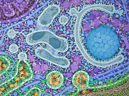
Don’t let the textbook diagram of a simplified two-dimensional cell fool you – within this tiny structure of life is a complex universe of molecular machinery that is continually being built, put into motion and eventually broken down.
Cells use the thousands of different proteins within them as tools to shape their internal environment. In this environment are specialized compartments known as organelles that carry out the cell’s functions. Two important organelles within cells are mitochondria and the endoplasmic reticulum, which produce energy and assemble proteins, respectively.

Since routine cellular activity generates toxic byproducts that can damage the cell, a disposal system is needed to degrade and recycle these molecules within cells. One of these processes is autophagy, a form of self-consumption cells use to eliminate and recycle abnormal or excess components, including proteins and organelles. Derived from Greek, the term literally translates to “self-eating.” In 2016, cell biologist Yoshinori Ohsumi won the Nobel Prize in Physiology or Medicine for his work on autophagy. Autophagy is essential for cellular health and longevity. When this process is not working well, it’s linked to several human diseases, including neurodegenerative and cardiovascular diseases and cancer.
We are researchers studying how autophagy is activated in cells. In our recently published research, we examined two key regulators of this process and identified a unique role one of them plays in degrading mitochondria that may serve as a potential target to treat certain diseases.
Autophagy and human disease
The connection between autophagy and disease is complex and not well understood.
For instance, autophagy appears to play a paradoxical role in cancer. On one hand, some studies have shown that because this process suppresses tumors by eliminating potentially harmful material, reduced or impaired autophagy can turn a cell cancerous. On the other hand, activating autophagy after a tumor has formed can promote cancer by helping it adapt and survive, potentially leading to treatment resistance.
These findings suggest that it is especially important to understand the precise steps and timing of autophagy when it comes to targeting this process as a cancer treatment strategy. Researchers are evaluating the anticancer effects of two malaria drugs, chloroquine and hydroxychloroquine, that block the final steps of autophagy. So far, they have varying efficacy depending on cancer type and stage.
Dysfunctional autophagy also plays an important role in most neurodegenerative diseases. The aggregation of abnormal proteins in brain cells are common features in Alzheimer’s disease, Parkinson’s disease, Huntington’s disease and ALS. Some scientists believe that the accumulation of these proteins is due at least in part to a decline in their degradation through autophagy.
Autophagy is also important for heart health. Researchers have found that autophagy in the heart declines with age and contributes to cardiovascular disease. Decreased autophagy in cardiac muscle cells results in accumulating cellular garbage that can affect their ability to contract and even cause their death. With fewer cells and less contraction, the buildup of toxic material in cardiac muscle cells can ultimately lead to heart failure.
Breaking down mitochondria with mitophagy
For autophagy to be efficient, it needs to specifically get rid of only damaged proteins or organelles within the cell. Uncontrolled degradation would deprive a cell of its basic needs.
This is particularly true for mitochondria, as cells rely on them for much of their energy production. Our team has been very interested in how cells ensure that autophagy of mitochondria, also known as mitophagy, eliminates only dysfunctional mitochondria while sparing the healthy parts of the cell. Dysfunctional mitophagy has been linked to cancer, neurodegeneration and cardiovascular disease, among other diseases.
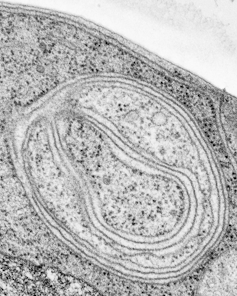
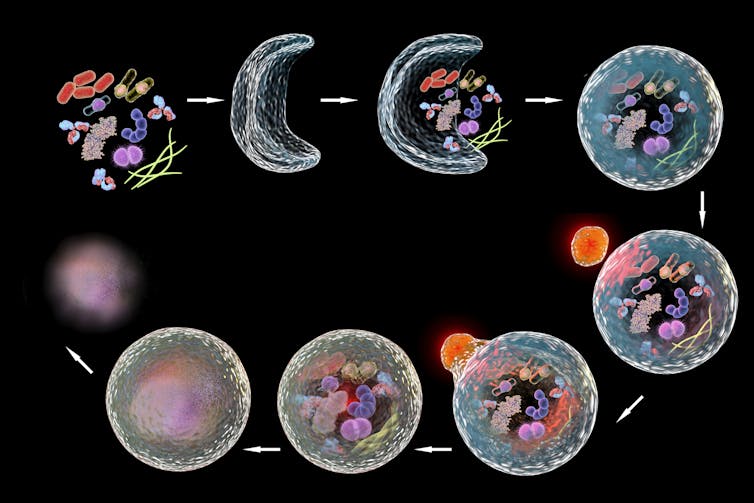
The process of autophagy starts when the cell begins to form a membrane near damaged proteins or organelles. This membrane will expand into a vesicle, or sac, known as an autophagosome, that engulfs the damaged material. It will then fuse with another internal cell structure full of acid called a lysosome that helps degrade its cargo.

Beclin1 is a protein known to promote the formation of autophagosomes in cells. However, its role in mitophagy is controversial, in part because very little is known about its close relative Beclin2. We wanted to disentangle the functions of these two proteins and determine their role in mitophagy. To do this, we used mouse and human cell models to examine how the presence or absence of these two proteins affected autophagy.
We discovered that activating a region unique to Beclin1 enables it to promote autophagosome formation next to dysfunctional mitochondria, facilitating their degradation in human cells. Because a similar region isn’t found in Beclin2, this meant that only Beclin1 may be essential for mitophagy.
Interestingly, we also observed Beclin1 at discrete points of contact between mitochondria and endoplasmic reticulum during mitophagy. This supports emerging research suggesting that physical interactions between these organelles facilitate the transfer of certain molecules needed to make autophagosomes. Our work indicates that only Beclin1 promotes engulfment of damaged mitochondria at these sites. Beclin2 may perform a different role in autophagy in other conditions.
Targeting autophagy for treatments
Autophagy represents a potential treatment target for many different diseases. Our team is currently studying how autophagy contributes to protein aggregation and mitochondrial dysfunction in the heart, and we are working to develop new tools to measure this process in cell and animal models.
However, therapeutic strategies to regulate autophagy is complicated by the fact that it is a complex multi-step process that involves many different proteins. Some diseases may require targeting the early steps of autophagsosome formation, while others may require focusing on when they fuse with lysosomes. Furthermore, different disease states may benefit from either autophagy activation or inhibition. More work needs to be done to identify all of the specific proteins that regulate each step of the autophagy pathway and how cells finetune this process in both health and disease.
We believe that helping cells better harness the power of autophagy in a complex molecular universe can train them to follow the three Rs – reduce, reuse, recycle – to promote health and longevity.
Åsa Gustafsson receives funding from NIH.
Justin Quiles receives funding from The American Heart Association.
Read These Next
Drug company ads are easy to blame for misleading patients and raising costs, but research shows the
Officials and policymakers say direct-to-consumer drug advertising encourages patients to seek treatments…
Tiny recording backpacks reveal bats’ surprising hunting strategy
By listening in on their nightly hunts, scientists discovered that small, fringe-lipped bats are unexpectedly…
There aren’t enough geriatricians – here’s how older adults can still get the right care
A few simple strategies can help older adults convey their needs to their health care provider.


